
Explore an animal cell
How do structures inside a cell work together to send a protein outside?
Explore seven structures from the whole cell to its organelles, then follow one secreted protein step by step. Plain-language explanations connect each location to its role.
Learning goals
- Identify the membrane, cytoplasm, nucleus, and four other structures in a representative animal cell.
- Explain the different roles of ribosomes, ER, Golgi apparatus, and mitochondria in plain language.
- Trace one secreted protein in order and explain which case the route represents.
Before you begin
- Know that cells are basic units of living structure and function.
- Use arrows to show movement from one location to another.
Explore an animal cell
Choose a structure name for an explanation. On larger screens, you can also choose image markers. Start 3D to rotate, pinch or scroll to zoom, and use names for close-ups.
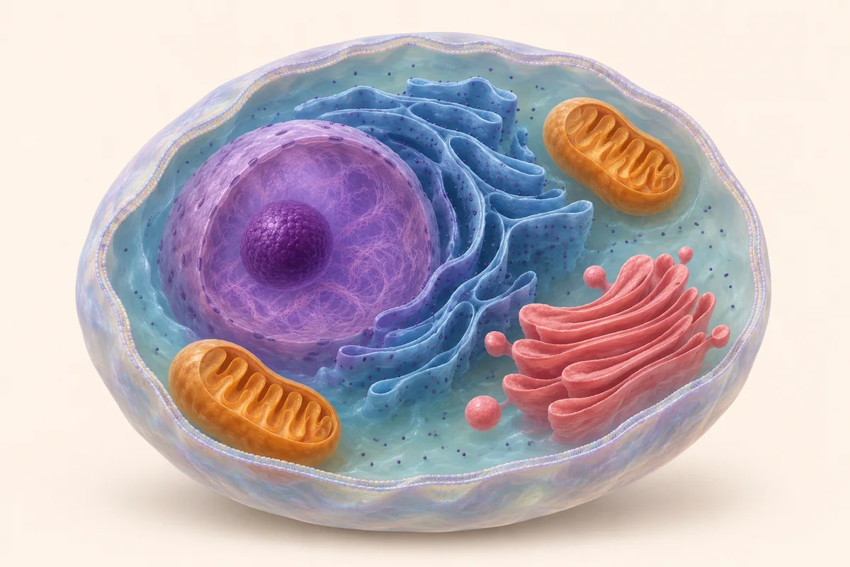
The flat view is ready. Choose a structure or play the route.
Reduced motion is on: continuous playback and camera animation are off. Use Previous step and Next step.
The offline lesson includes the flat interaction and all explanations. Open the online lesson for 3D.
Choose a structure to discover its job
Cell membrane
In everyday words
Think of the membrane as a selective doorway. It separates the cell from its surroundings and helps control which substances enter or leave.
Keep this in mind:The doorway is an analogy. Substances cross membranes in different ways; not everything passes freely. The large cutaway lets you see inside and is not a real opening in the membrane.
Cytoplasm
In everyday words
The cytoplasm is the region inside the cell membrane but outside the nucleus. It includes the fluid, many organelles, and the supporting cytoskeleton.
Keep this in mind:The gel-like fluid is called cytosol; it is only one part of the cytoplasm. This diagram omits much of the cytoskeleton and many other structures.
Nucleus
In everyday words
Think of the nucleus as a store for the main instruction set. It contains most of the cell’s DNA, whose information supports many cell activities.
Keep this in mind:Not all DNA is in the nucleus: mitochondria have their own DNA too. The store-room analogy does not imply that the nucleus thinks or that proteins are made on ribosomes inside it.
Mitochondria
In everyday words
Think of mitochondria as energy-conversion stations. They take part in cellular respiration, transferring some chemical energy from nutrients into ATP that the cell can use.
Keep this in mind:Mitochondria do not create energy from nothing and are not the only place where a cell produces ATP. Their inner-membrane folds are cristae; only a few enlarged folds are shown here.
Ribosomes
In everyday words
Ribosomes are like tiny assembly benches. They use genetic instructions to join amino acids into proteins. Some are free in the cytosol; others attach to rough endoplasmic reticulum.
Keep this in mind:The dots are greatly enlarged so you can find them. Ribosomes are not enclosed by a membrane. Their attachment to rough ER does not mean the ER alone builds proteins.
Endoplasmic reticulum (rough ER shown)
In everyday words
Rough ER is a connected membrane network with ribosomes attached. Certain proteins destined for secretion enter it, where folding and processing begin.
Keep this in mind:“Rough” refers to attached ribosomes, not spikes on the membrane. Smooth ER lacks these attached ribosomes and has other functions; it is not fully represented here.
Golgi apparatus
In everyday words
The Golgi apparatus is a stack of flattened sacs that receives, processes, and sorts cargo. Transport vesicles arrive from the ER; some cargo is further modified and sent to its destination.
Keep this in mind:Cargo arrives at the receiving (cis) face and leaves from the shipping (trans) face. Not every protein passes through the Golgi, and not all Golgi cargo is sent outside the cell.
How to read this model
Think of a cell as a tiny workspace: a membrane surrounds structures that work together. The guide is an AI-assisted artistic illustration checked for the structures taught here; the 3D model is independently built in code. We cut away the front to show the interior. A real cell has no such opening. Colours, sizes and numbers are not microscopy results.
Follow one protein destined for secretion
This route follows one protein being sent outside the cell. The yellow marker shows its location; later transport involves vesicles. Not all proteins follow this route.
We follow one protein destined for secretion. A ribosome attached to rough ER builds it; the protein enters the ER and begins folding and processing.
A transport vesicle carries this cargo from the ER to the Golgi’s receiving face. The yellow marker represents the tracked cargo, not its real size or speed.
Cargo arrives at the cis face, is further processed and sorted in the Golgi, and leaves from the trans face. A single simplified path represents this stage.
For our selected secreted protein, a secretory vesicle carries it from the Golgi toward the cell membrane. Other cargo may have different destinations.
The secretory vesicle fuses with the cell membrane, releasing its protein cargo outside by exocytosis. This does not leave a permanent hole in the membrane.
Read all structure explanations
Cell membrane
Think of the membrane as a selective doorway. It separates the cell from its surroundings and helps control which substances enter or leave.
The doorway is an analogy. Substances cross membranes in different ways; not everything passes freely. The large cutaway lets you see inside and is not a real opening in the membrane.
Cytoplasm
The cytoplasm is the region inside the cell membrane but outside the nucleus. It includes the fluid, many organelles, and the supporting cytoskeleton.
The gel-like fluid is called cytosol; it is only one part of the cytoplasm. This diagram omits much of the cytoskeleton and many other structures.
Nucleus
Think of the nucleus as a store for the main instruction set. It contains most of the cell’s DNA, whose information supports many cell activities.
Not all DNA is in the nucleus: mitochondria have their own DNA too. The store-room analogy does not imply that the nucleus thinks or that proteins are made on ribosomes inside it.
Mitochondria
Think of mitochondria as energy-conversion stations. They take part in cellular respiration, transferring some chemical energy from nutrients into ATP that the cell can use.
Mitochondria do not create energy from nothing and are not the only place where a cell produces ATP. Their inner-membrane folds are cristae; only a few enlarged folds are shown here.
Ribosomes
Ribosomes are like tiny assembly benches. They use genetic instructions to join amino acids into proteins. Some are free in the cytosol; others attach to rough endoplasmic reticulum.
The dots are greatly enlarged so you can find them. Ribosomes are not enclosed by a membrane. Their attachment to rough ER does not mean the ER alone builds proteins.
Endoplasmic reticulum (rough ER shown)
Rough ER is a connected membrane network with ribosomes attached. Certain proteins destined for secretion enter it, where folding and processing begin.
“Rough” refers to attached ribosomes, not spikes on the membrane. Smooth ER lacks these attached ribosomes and has other functions; it is not fully represented here.
Golgi apparatus
The Golgi apparatus is a stack of flattened sacs that receives, processes, and sorts cargo. Transport vesicles arrive from the ER; some cargo is further modified and sent to its destination.
Cargo arrives at the receiving (cis) face and leaves from the shipping (trans) face. Not every protein passes through the Golgi, and not all Golgi cargo is sent outside the cell.
One quick model check
Observe, then explain
Work through four steps. Each button moves the interactive model to the matching observation state.
-
The selectively permeable membrane separates the cell from its surroundings. The cytoplasm between the membrane and nucleus includes cytosol, organelles, and the cytoskeleton, rather than just water.
Look for: Select “Cell membrane”, then “Cytoplasm”. Explain how the outer boundary differs from the region inside.
-
The nucleus stores most of the cell’s DNA, and ribosomes use genetic instructions to make proteins. The nucleus is not a tiny person giving orders: cell activities depend on interacting structures and chemical processes.
Look for: Select the nucleus and ribosomes. Explain the difference between storing genetic information and making a protein.
-
Mitochondria take part in cellular respiration, transferring some chemical energy from nutrients into ATP. Many cell processes use ATP, so “energy-conversion station” is a better analogy than creating energy from nothing.
Look for: Select mitochondria and find the inner folds. Explain where the energy comes from instead of memorising “powerhouse”.
-
For the secreted protein selected here, an ER-bound ribosome builds it. It enters the ER, travels in a vesicle to the Golgi, then leaves in a secretory vesicle that fuses with the membrane to release it outside.
Look for: Step through the route, then describe it in order yourself. Explain why mitochondria are not a delivery stop on this route.
Build an explanation
Connect each location with a role
The aim is more than memorising seven names. Find the membrane, distinguish the nucleus from the cytoplasm, then relate structures to their roles. Cytoplasm includes several structures, and organelles do not work as isolated little rooms.
Assembly and sorting are different jobs
Ribosomes make proteins. Rough ER receives and begins processing some of them, and the Golgi continues processing, sorting, and shipping. These analogies describe different roles; real molecular interactions do the work, without workers adding parcel labels.
One route does not cover all proteins
This lesson chooses one protein secreted through the ER and Golgi. Many proteins work inside the cell and do not follow this complete route. Even cargo passing through the Golgi may be sent to another structure rather than outside.
Connect with respiration and diffusion
Ventilation, gas diffusion across membranes, and cellular respiration are different processes. The breathing-and-circulation lesson follows oxygen to tissues; this lesson introduces mitochondria involved in cellular respiration. Selective membrane transport includes more than simple diffusion.
Printable worksheet
Understand the concept here, then work independently on the A4 sheets. Practice, calculations and investigation records are included together. Reference answers open only in the parent dialog.
Biology
Explore an animal cell
How do structures inside a cell work together to send a protein outside?
- Which statement best describes the cell membrane?
A. It lets every substance pass freely. B. It separates the cell from its surroundings and selectively controls exchange. C. Its main role is storing most of the cell’s DNA.
- In a nucleated animal cell like this model, which statement about DNA is correct?
A. Most is in the nucleus; mitochondria also have their own DNA. B. All of it is only in the nucleus. C. It is found only in the Golgi apparatus.
- Consider only the protein secreted through ER and Golgi in this lesson. Which simplified route is correct?
A. Mitochondria → nucleus → cell membrane B. Golgi → nucleus → rough ER C. Rough ER → transport vesicle → Golgi → secretory vesicle → cell membrane
- Someone says, “Mitochondria create energy from nothing.” Which correction is accurate?
A. They transfer some chemical energy from nutrients into ATP for cell use. B. Their only job is sending proteins outside the cell. C. They move all DNA to the cell membrane.